It’s the year’s end, and you have been busy working late nights and rushing for the year-end dateline before Christmas or Chinese New Year. You must rush home and pack for the huge family trip you have planned for the past year. As you head back, you suddenly feel the beginning of an intermittent squeezing pain over the back. This pain rapidly increases in intensity, and as you reach home, you are bathed in sweat, feel nauseous and barely able to stand. You cannot find a position that makes the pain slightly bearable. Soon, you are lying on the floor, wringing and screaming in pain, experiencing the worst possible pain in your life.
The intensity of the pain is surpassed only by the fear of what is happening to your body. You vomit out your lunch, not knowing whether it is going to be the last meal you take as you head straight to the nearest emergency department. And that is the most common presentation of renal colic.
How Common Are Stones?
Kidney stones are a common condition in Singapore as we are located along the stone belt, where kidney stones are more prevalent (common). Different countries have different prevalences of kidney stone disease, which can be as high as 16% in Southeast Asia and nearly 20% in Saudi Arabia. The climate influences the incidence of kidney stone, fluid intake, and the local regional diet of choice. Not surprisingly, kidney stones are not as common in temperate countries, but they are common in regional countries like Indonesia, India, Thailand, Myanmar, and Malaysia.
Men are twice as likely as women to have kidney stones. Over 70% of kidney stone occur between the 20- to 50-year-old age group. There is a saying; “once a kidney stone former, always a kidney stone former”. More than 50% of patients with kidney stones will form kidney stones within five years if the causative factors are not removed.
Symptoms
Acute renal colic is a severe form of pain that occurs when a kidney stone dislodges itself from the kidney and drops into the ureter. As the ureter is a narrow passageway for urine to flow from the kidney into the bladder, kidney stones can lodge in the ureter and stop urine flow. The kidney distends itself as urine is continuously made, and the stretching of the kidney causes a pain score of 10/10 and is often described as worse than child-bearing pain. Renal colic usually requires potent analgesia (painkillers) injection.
Without treatment, the urine flow is obstructed, and obstructions affect kidney function. Urosepsis occurs when the obstructed urine becomes infected and can be a life-threatening condition. Even if infection does not happen, long-term obstruction of the kidney leads to the destruction of the obstructed kidney. I have patients as young as 20s having lost one of their kidneys due to previously undiagnosed kidney stone diseases.
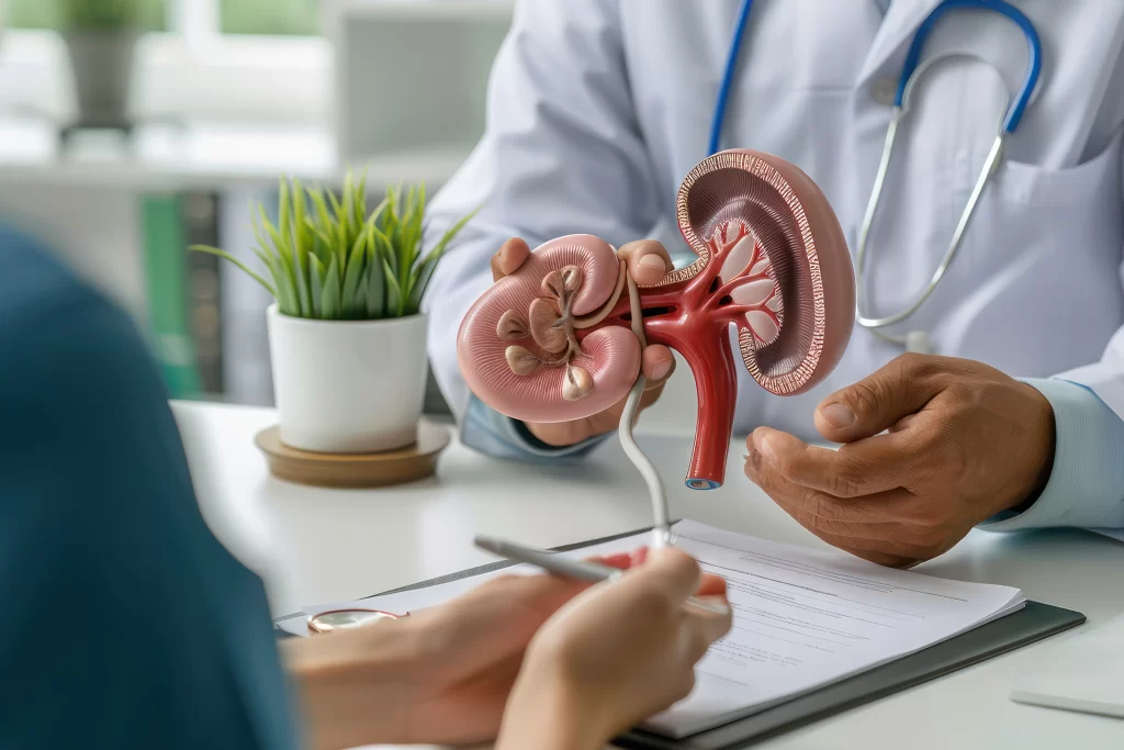
Diagnosis
Kidney stones can be diagnosed through a combination of history taking, physical examination, imaging, and laboratory/urine tests. There are a variety of imaging techniques that are used to diagnose kidney stones. The ‘gold standard’ modality is the Computated Tomography (CT). CT can detect more than 90% of all types of kidney stones. Other modalities that can be useful include abdominal X-ray or ultrasound, which can be used to image the kidney and bladder effectively. However, the sensitivity of ultrasound/X-ray is lower than that of CT when imaging small kidney stones. In addition, CT can image the ureters, which the ultrasound cannot. For most people, CT is the image of choice if available.
Once a diagnosis is made, treatment goals would be to:
- 1. Control the pain and/or symptoms,
- 2. Unblock the obstruction and preserve kidney function.
Depending on the availability of services, the pain can be managed either via a hospital admission with painkiller injections for severe pain or discharge back home with oral medication. Regardless, you must see a urologist as soon as possible for goal number 2.
Treatment: Unblocking the Obstruction
There are generally three main ways to unlock the obstruction.
Conservative Management of Kidney Stones
Medications and advice on analgesia control may be given to help kidney stone passage. Conservative management of renal colic is only possible if the obstructing kidney stone is 4mm or less, as they have a high chance of spontaneous passage within four weeks. 90% of kidney stones 4mm or less will eventually passed out, but do note that the four weeks may be pocketed by sudden acute renal colic attack any time requiring a trip to the emergency department repeatedly while awaiting the transit of kidney stone through the ureter. Beyond four weeks, it is recommended that intervention is performed to remove the obstruction and prevent scarring from prolonged obstruction.
Extracorporeal Shockwave Lithotripsy (ESWL)
ESWL uses energy generated from shockwaves to compress and expand small air pockets within kidney stones repeatedly. This procedure requires only a light sedation of the patient to prevent movement during the procedure. This procedure can be performed as a day surgery and discharged home. The procedure’s benefits are that there is no invasive component, and general anaesthesia is avoided. The flip side of the procedure is that it is not suitable for obese patients, pregnant patients, or patients who have kidney stones that are not visible. It is important to note that ESWL breaks up the kidney stone, and how it moves down the ureter is unpredictable. Steinstrass occurs when the leading kidney stone gets lodged downstream after the ESWL, possibly leading to a renal colic attack. The efficacy of this modality is about 75% for small kidney stones less than 10mm and decreases as the kidney stone size increases.
Ureteroscopy and Laser Lithotripsy
This procedure is considered the ‘gold standard’ for unblocking obstruction as it can handle various kidney stone hardness, size, location, patient size, and kidney stone fragments removed by the surgeon. In this procedure, the surgeon slips a tiny instrument into the bladder and up along the ureter until the kidney stone is located. A laser fragments the kidney stone into small fragments and relieves the obstruction. The kidney stone fragments are then removed piece by piece to ensure kidney stones are not left behind. In addition, a double J ureteral stent is placed in the ureter to ensure the passage of urine from the kidney to the bladder while allowing the ureter to heal after surgery. It can handle large kidney stones regardless of the patient’s location and habitus. Patients with anti-platelet medication can also undergo this procedure as it is generally safe in experienced hands. The downside of the modality is that a general anaesthetic is required, but it is more than adequate and benefits from high stone-free rates and preventing severe infection.
The ureteric stent is removed in a simple, short procedure a few days later, and you will be kidney stone-free! Remember that if you do not modify your risk factors, you are likely to have kidney stones again. Hydration is a key component of prevention along with a healthy balanced diet. We will go through certain basic yet often overlooked principles of hydration in another article.