
As you complete your annual health screening, ultrasound of the kidney reveals a cystic lesion or small renal mass. What do you do? Renal masses are commonly identified during imaging studies performed for unrelated conditions. These abnormalities can range from benign to malignant, and understanding their nature is critical for effective management.
The Bosniak classification system is commonly used in evaluating cystic kidney lesions. The classification provides a standardized approach and categorizes lesions based on radiological features, enabling accurate risk assessment for malignancy.
Regular follow-up and imaging are essential for accurate diagnosis and long term management to ensure the character of the lesion remains the same.
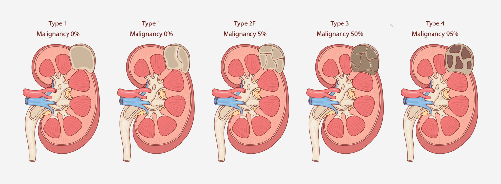
Simple cysts characterized by thin walls, no septa, calcifications, or solid components. These are water-density lesions that do not enhance with contrast and are universally considered benign. No follow-up is required.
Slightly more complex cysts that might have thin septa or fine calcifications. High-attenuation lesions less than 3 cm with sharp margins also fall under this category. These are benign and typically do not require follow-up.
Intermediate-risk cysts that may contain more septa, minimal wall thickening, or nodular calcifications. They show no soft-tissue enhancement but require periodic monitoring with imaging to confirm stability.
Indeterminate lesions with irregular walls or septa that enhance with contrast. These masses carry a 50% risk of malignancy and generally require surgical exploration.
Clearly malignant lesions containing enhancing soft-tissue components. These have an 80–90% risk of cancer and necessitate surgical removal.
Solid renal masses can raise concerns due to their potential for malignancy. However, there are several benign conditions mimic the presentation of malignant tumors, including:
Constituting about 5% of solid renal tumors, oncocytomas are non-cancerous growths that originate from the intercalated cells of the collecting ducts. Though benign, they are challenging to distinguish from renal cell carcinoma (RCC) without histopathological analysis. Oncocytomas often present with a central stellate scar on imaging and have a characteristic mahogany brown color on pathology.
These benign tumors are composed of fat, smooth muscle, and thick-walled blood vessels. They frequently occur sporadically but may also be associated with tuberous sclerosis complex (TSC). Larger AMLs (>4 cm) are prone to spontaneous hemorrhage and may present with acute pain or hematuria. Management includes embolization, nephron-sparing surgery, or mTOR inhibitors in select cases.
Some benign renal masses are associated with genetic syndromes, underscoring the importance of family history and genetic counseling in diagnosis:
This condition is characterized by bilateral renal cysts and complications such as hypertension, kidney failure, and extrarenal manifestations (e.g., liver cysts, intracranial aneurysms). Management focuses on blood pressure control, symptomatic treatment, and renal replacement therapy when necessary.
An autosomal dominant disorder that affects multiple systems, TSC is linked to AMLs and other renal manifestations. Patients may also exhibit neurological symptoms, skin lesions, and pulmonary complications. Early diagnosis and tailored management are crucial for improving outcomes.
If you suspect a renal mass or have been diagnosed with one, consult Dr Jay Lim for a tailored care plan.
Advanced imaging play a pivotal role in diagnosing and managing renal masses. Common ways to look at the kidney include:

Biopsy may be required for lesions where imaging findings are inconclusive, especially when distinguishing benign from malignant masses.
Biopsy may be required for lesions where imaging findings are inconclusive, especially when distinguishing benign from malignant masses.
Management of benign renal masses varies based on their type, size, symptoms, and associated risks:
Advancements in imaging and easy availability of imaging allows for a better understanding of renal mass biology, most benign renal masses can be effectively managed without the need for surgery. Regular follow-up with imaging is crucial for intermediate-risk lesions to detect changes early and guide treatment decisions.
If you suspect a renal mass or have been diagnosed with one, consult Dr Jay Lim for a tailored care plan.
Our friendly team at Urocare is looking forward to serving you.
For urgent enquiries and appointment requests, please call the clinic directly.
Please note that UroCare will be operating on a half day on 16th, and will be closed on 17th and 18th.
We will resume normal operating hours on 19th.
Thank you for your understanding.